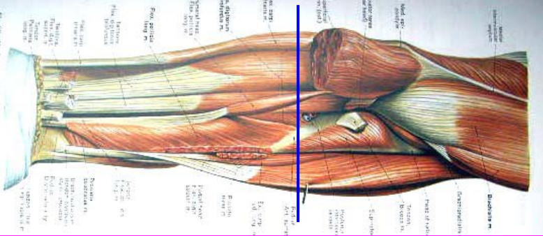
1. A long volar flap and a short dorsal flap were made, with a length ratio of 2:1.
The incision began at 1.3cm distal to the radial styloid process, extended to the distal end, and then curved to the proximal end, ending at 1.3cm distal to the ulna styloid process. (In order to preserve the length of limbs, non-standard flaps can be made if necessary)
2. The flap was detached to the proximal side of the planned osteotomy plane to expose the underlying soft tissue
3. Pull and cut the flexor and extensor tendons distally and allow them to retract into the forearm.
4. Dissociate the flexor and extensor carpus tendons from the attachment point and flip them to the proximal side of the osteotomy plane.
5. Locate the terminal branches of the positive and radial nerves, ulnar nerves and radial nerves, pull them to the distal end, and cut them with a sharp knife.
6. Proximal to the expected osteotomy plane, the radial and ulnar arteries were clamped, ligated, and severed.
7. Transection the carpal bone L and file the edges.
8. Select the same position on the residual carpal bone as the attachment point of the normal tendon, and fix the flexor carpal tendon and extensor carpal tendon to retain the active motion of the wrist joint.
9. Subcutaneous tissue and skin were sutured for drainage.

Wrist amputation differs from transcarpal amputation in the following cases
1. Annular cutting of wrist joint capsule
2. Remove the radial neck process and ulna styloid process, and flatten the residual end.
Attention should be paid to avoid damage to the radioulnar joint and triangular fibrous cartilage, so as to preserve the pronation and supination functions of the forearm, and avoid joint pain.
Forearm amputation
The characteristics of
1. Thin skin on the distal forearm;
2. The deep tissue of the distal forearm is basically composed of tendons and fascia with relatively poor blood transport.
Therefore, amputation at 1/3 of the forearm is preferable when there is severe disruption of blood circulation to the forearm.
Amputation of the distal forearm
1. Make anteroposterior isometric flaps from the proximal end of the anticipated osteotomy plane.
2. Flip the flap, subcutaneous tissue, and deep fascia proximal to the osteotomy plane.
3. Handle blood vessels and nerves.
4. The distal end of the osteotomy plane transects the muscular abdomen of the forearm.
5. Cut the ulnar and radius horizontally and file the bone ends flat.
6. Suture and drainage.
Myoplasty is performed for the needs of modern prosthetics.
1. After lifting the skin and deep fascia of appropriate length, the superficial flexor tendon is constructed into a muscle flap that is long enough to bypass the stump of the osteotomy to reach the deep dorsal fascia.
2. Transection other soft tissues at the osteotomy plane.
3. Osteotomy and filing were performed, and the flap was turned dorsally over the dorsal muscle and sutured with the deep fascia.
Note: In order to prevent residual bloatiness, the entire volar muscle should never be made into muscle flap.
Proximal 1/3 amputation of the forearm
1. The skin condition is good, do isometric skin flap before and after; If conditions are not good, in order to preserve the length, if necessary, do informal flaps.
2. Invert the flap and deep fascia proximal to the osteotomy plane.
3. Handle blood vessels and nerves.
4. Transection muscle abdomen at the distal end of the articular plane. If the stump is above the biceps insertion, the distal biceps should be removed as described by Blair and Morris. This will increase the length of the functional stump and facilitate the fitting of the prosthesis.
Amputation of the elbow
1. The anterior and posterior stage flaps were made, starting from the inner and outer condyles of the humerus, extending the posterior flaps to 25cm distal to the olecranon of the ulna, and extending the anterior flaps to the distal side of the biceps tendon insertion.
2. Turn the flap up, starting from the elbows in deep tissue separation, membrane processes in turn two tendons, flexor tendon always start, brachial artery, the median nerve, ulnar nerve, and then from the radial stripping biceps tendon of check point, the brachial muscle, brachial radial groove in the separation between muscle, cut off the radial nerve, in 63 cm distal joint line, cross sectional extensor group nearly end flip up.
3. Near the olecranon of the ulna, the triceps tendon and the posterior fascia were incised, and the joint capsule was then incised to remove the forearm and complete the dissociation.
4. Triceps brachii tendon was moved forward and sutured with brachii tendon and biceps brachii tendon. Part of extensor muscle group left on the external epicondyle of humerus was pruned into thin muscle flap, which was pulled inward and sutured with the stump from the upper condyle of the internal humerus.
5. Periosteum and muscle flap covering all bony prominences and exposed tendons of the humeral stump.
6. Suture and drainage
Upper arm amputation
Amputation at any level from the supracondyle of the humerus to the axillary fold. The characteristics of
1. The prosthesis of an above-elbow amputee must have an internal locking device and an elbow turntable. The locking device of the elbow should be 3.8cm distal to the receiving cavity. Therefore, when performing an above-elbow amputation, the osteotomy level should be at least 3.8cm from the elbow to allow room for the installation of the device.
2. The humeral head is preserved -- the normal shape of the shoulder joint is preserved so that the socket of the prosthesis is easy to grasp and the prosthesis is more stable.

 Thigh Series
 Lower Leg Series
 Product Video
 Group Propaganda Video